- TEL:
- +81-3-5462-4831
- FAX:
- +81-3-5462-4835
※9:00-17:40 Mon.-Fri. (JST)


|
The inner wall of PrimeSurface products are coated with our unique ultra hydrophilic polymer, which prevents a cell from adhering to the plastic surface induces a spontaneous cell spheroid formation in equal size and shape. |
For Stem Cell Research:
Induction of ES, iPS and mesenchymal stem cells differentiation by embryoid body formation.
For Drug Screening Research & Development:
An ideal product for drug response studies. Three dimension spheroid models are more physiologically relevant than two dimension monolayer model.
| Cat. No | Product | Description | Culture Area | Volume | Qty/Pk | Qty/Cs |
|---|---|---|---|---|---|---|
| MS-90350 | PrimeSurface Dish 35 | 35 Φ × 14(H)mm | 9 cm2 | - | 5 | 50 |
| MS-90600 | PrimeSurface Dish 60 | 60 Φ × 15(H)mm | 21 cm2 | - | 10 | 120 |
| MS-90900 | PrimeSurface Dish 90 | 90 Φ × 20(H)mm | 57 cm2 | - | 10 | 50 |
| MS-90240 | PrimeSurface Plate 24F | 24wells, Flat | 1.8 cm2 | 3.4mL/well | 1 | 10 |
Remark
| Cat. No | Product | Wells | Bottom | Volume | Qty/Pk | Qty/Cs |
|---|---|---|---|---|---|---|
| MS-9384U | PrimeSurface 384U | 384 | U bottom | 0.1mL | 1 | 20 |
| MS-9384W | PrimeSurface 384U White Plate | 384 | U bottom | 0.1mL | 1 | 20 |
| MS-9096VZ | PrimeSurface 96V | 96 | V bottom | 0.3mL | 1 | 20 |
| MS-9096MZ | PrimeSurface 96M | 96 | Spindle bottom | 0.2mL | 1 | 20 |
| MS-9096UZ | PrimeSurface 96U | 96 | U bottom | 0.3mL | 1 | 20 |
| MS-9096WZ | PrimeSurface 96U White Plate | 96 | U bottom | 0.3mL | 1 | 20 |
Remark
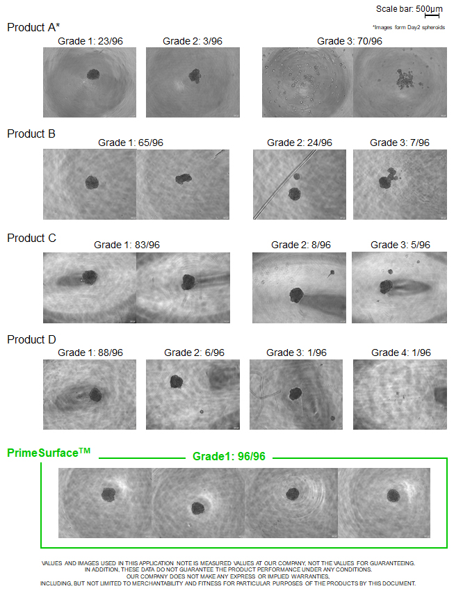
| Cell line | : | HepG2 |
|---|---|---|
| Seeding density | : | 1000 cells/well |
| Medium | : | DMEM+10%FBS |
| Culture period | : | 3 days |
| Plate | : | 96 well U plates for 3D culturing from different companies |
Classification based on microscopic images
| Manufacturer | Product | Grade 1 | Grade 2 | Grade 3 | Grade 4 |
|---|---|---|---|---|---|
| Company A | Product A | 23 | 3 | 70 | 0 |
| Company B | Product B | 65 | 27 | 4 | 0 |
| Company C | Product C | 83 | 8 | 5 | 0 |
| Company D | Product D | 88 | 6 | 1 | 1 |
| Company E | Product E | 95 | 1 | 0 | 0 |
| S-BIO Sumitomo Bakelite Co., Ltd. |
PrimeSurface™ | 96 | 0 | 0 | 0 |
Remark
※PrimeSurface™ has the advantage of supporting single well single spheroid formation
| Product A** (n=23) |
Product B (n=65) |
Product C (n=83) |
Product D (n=88) |
Product E (n=95) |
PrimeSurface™ (n=96) |
|
|---|---|---|---|---|---|---|
| Ave (μm) | 225.4 | 244.4 | 254.8 | 238.7 | 261.6 | 240.4 |
| SD | 16.5 | 11.3 | 11.7 | 10.1 | 9.4 | 7.3 |
Remark

※PrimeSurface™ has the advantage of supporting uniform spheroids formation
【PrimeSurface™ is a great tool to obtain highly reproductive data
by providing uniform spheroids in size】

Regenova is a novel robotic system that facilitates the fabrication of three- dimensional cellular structures by placing cellular spheroids in fine needle arrays according to pre-designed 3D data. Followings are examples of such fabrications using S-BIO's PrimeSurface™ 96U plate and Bio 3D Printer, Regenova (Cyfuse Biomedical K.K.).; neural 3D tissue and 3D tissue with mesenchymal stem cells.
| Cell sources | : | hiPSC derived neural progenitor cells |
|---|---|---|
| Quantity of cells | : | 4 X 10⁴ cells/well |
| Culture Medium | : | For neural cells |
| Maturation duration on Well Plate | : | 2 days |
| 3D structure of printed 3D tissue | : | printed with 3 X 3 X 2 |
| Quantity of spheroids used for 3D tissue | : | 18 spheroids |
| Maturation duration after 3D printing | : | Remove needles 9 days after 3D print |
| Cell sources | : | human adipose tissue derived mesenchymal stem cells (hADSC) |
|---|---|---|
| Quantity of cells | : | 5 X 10³ cells/well |
| Culture Medium | : | For MSC |
| Maturation duration on Well Plate | : | 2 days |
| 3D structure of printed 3D tissue | : | Circle shape with 48 spheroids x 10 layers |
| Quantity of spheroids used for 3D tissue | : | 480 spheroids |
| Maturation duration after 3D printing | : | Remove needles 6 days after 3D print |
| Culture plate | : | PrimeSurface MS-9096V |
|---|---|---|
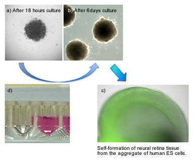
| Cell type | : | Human ES cells (KhES-1 strain) |
| Seeding density | : | 9,000 cells/well |
| Culture medium | : | GMEM+KSR+NEAA+2ME+ 20uM Y-27632 |
| Culture environment | : | 5%CO2, 37°C |

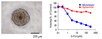
| Cell type | : | MCF-7 (Human breast cancer cell) |
|---|---|---|
| Anticancer drug | : | 5-Fluorouracil (5-FU) |

Nishio Lab., Department of Genome Biology, Kinki University School of Medicine
Please check the supplemental data for more detail example of application.

[Culture conditions]
| Culture plate | : | PrimeSurface MS-9096V |
|---|---|---|
| Cells | : | hiPS cell (201B7: Takahashi K et al. Cell, 2007 Nov 30;131(5):861-72, iPS Academia Japan, Inc.) |
| Seeding density | : | 9,000 cells/well |
| Culture medium | : | DMEM/F12 + 20% (v/v) KSR + 1% (v/v) NEAA + L-Glutamine (2mM) + Β-Mercaptethanol (80μM) + Y-27632(30μM) |
| Culture environment | : | 5%CO2, 37°C |
[Culture conditions]
| Seeding density | : | 1,000 cells/well |
|---|---|---|
| Culture medium | : | MEM+10FBS 100μL/well |
| Culture period | : | 48hours |
[Culture conditions]
| eding density | : | 1,000 cells/well |
|---|---|---|
| Culture medium | : | DMEM (Low Glucose) + 10%FBS 50μL/well |
| Culture period | : | 48hours |