- TEL:
- +81-3-5462-4831
- FAX:
- +81-3-5462-4835
※9:00-17:40 Mon.-Fri. (JST)


Tanaka, M., Chuaychob, S., Homme, M., Yamazaki Y., Lyu R., Yamashita K., Ae K., Matsumoto S., Kumegawa K.,
Maruyama R., Qu W., Miyagi Y., Yokokawa R. and Nakamura T.
Nat Commun 14, 1957 (2023). https://doi.org/10.1038/s41467-023-37049-z
Copyright: © 2023 by the authors. This article is an open access article distributed under the terms
and conditions of the Creative Commons Attribution (CC BY) license (https:// creativecommons.org/licenses/by/ 4.0/).
Tumor angiogenesis is one of the most important processes in the malignant progression and distant metastasis of cancer.
Alveolar soft part sarcoma (ASPS) is a rare cancer that typically occurs in young adults and is characterized by poor prognosis. While ASPS has low invasiveness at the primary site, it has a high propensity for metastasis. ASPS is characterized by a vascular-rich alveolar structure and its highly integrated vascular network is responsible for the frequent metastases. Understanding the mechanism of angiogenesis in ASPS is further expected to lead to the development of new treatment methods.
The fusion transcription factor ASPSCR1::TFE3 is necessary for tumor development in vivo. The group also created a model reflecting alveolar structure in vivo to evaluate angiogenic potential.
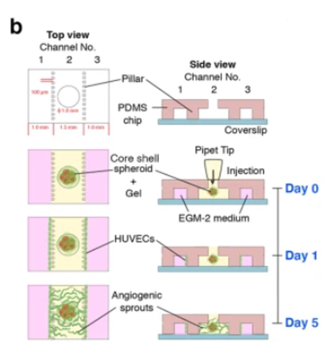
A core shell structure containing ASPS tumor cells covered with pericytes was formed using PrimeSurface™ and an innovative organ-on-a-chip system:
| 1. | 5.0 x 10⁴ cells/mL ASPS cells were cultured for 2 days in a PrimeSurface 96U plate. |
|---|---|
| 2. | 7.5 x 10⁴ cells/mL Pericytes were added to the ASPS cell spheroid core for shell formation |
| 3. | The formed spheroids with a core shell structure were then introduced into a microfluidic device, as shown in the figure (Day 0). |
| 4. | HUVEC cells were seeded into the different channels (Day 1) and co-cultured with the spheroids. Vascular formation was evaluated 4 days later (Day 5). |
Results: significant HUVEC sprouting toward the tumor spheroids of ASPS cells and Pericytes was observed during co-culturing.

The structure of spheroid formation on the three-channel microfluidic device
| Cat # | Product name | Well | Color | Bottom design | Well Vol | Package |
|---|---|---|---|---|---|---|
| MS-9096UZ | PrimeSurface™ 96U | 96 | Transparent | U bottom | 300 μL | Individual packaging 20 plates per case |
Remark