製品紹介
 |
|
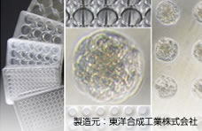
Cell-able® は、難しい操作や機器は一切不要で、通常のプレートに播種するように細胞を播種するだけで、1ウェルの中に均一なサイズの多数のスフェロイドを形成させることが可能です。
またスフェロイドが底面に接着しているため、蛍光プローブ染色、免疫染色などを容易に行うことができます。
肝毒性の蛍光染色イメージングと解析
ImageXpress® Micro XL System and MetaXpress® Softwear (Molecular Devices) によるイメージングと解析

Cell-able® における肝細胞とフィーダー細胞の共培養
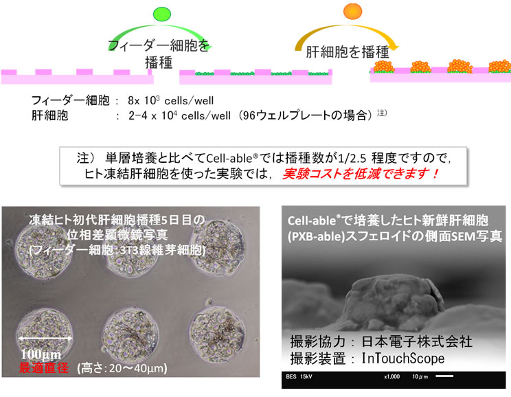
Cell-able®上で培養した新鮮ヒト肝細胞スフェロイド (3T3 Swiss albino との共培養)
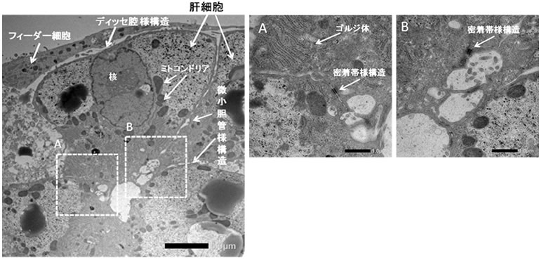
キメラマウス由来ヒト新鮮肝細胞(PXB-cells®, フェニックスバイオ)をCell-able® に播種後10日目の電子顕微鏡写真。肝微小構造の再構築が観察された。(データ監修: 旭川医科大学 医学部 病理学講座 腫瘍病理分野 西川祐司教授)
特長
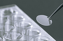
- 細胞接着面がアレイ状に配置されたマルチウェルプレート
- 一般的な培養器と同じようにプレートに細胞を播種するだけで、簡単にスフェロイドが形成される
- 形成されるスフェロイドサイズはほぼ均一であるため、良好な試験再現性が得られる
- 初代培養器肝細胞や株化がん細胞および患者由来のがん細胞など、接着性の細胞がスフェロイドを形成
Cell-able® でのスフェロイド形成
- Cell-able® の培養表面は円形の細胞接着領域が多数パターニングされています。接着領域以外は特殊なポリマーでコートされており、細胞が接着することは出来ません。
- Cell-able® に細胞を播種すると、細胞は自ら細胞接着領域に集まって接着します。増殖する細胞は、その場で増殖しスフェロイドを形成します。初代肝細胞のように増殖しない細胞の場合は、播種する細胞数を増やすことにより、スフェロイドが形成されます。
用途
創薬研究・バイオ研究
- 薬剤の肝代謝・毒性評価
- 抗がん剤のHCS (High Content Screening)
- マルチカラーイメージング解析を用いた心毒性評価
仕様・価格
| シリーズ | 品番 | 品名 | 内容 | 数量 | 参考価格(円) |
|---|---|---|---|---|---|
| Cell-able® プレート |
BS-9096CK | BP-96-R800 | 96 well plate, black wall, clear bottom, 800 circles/well |
1枚 | 28,000 |
- プレート有効期限: 製造後20ヶ月(2~8℃保存)(冷蔵輸送)
- 参考価格(本体価格・税抜)
- プレートは放射線滅菌済み
- 本製品は研究用 です。臨床用途には使用できません。
- 納期: 10日前後
- 現在欠品中ですが、次回2025年4月の入荷をもって販売終了。数量につきましては別途ご連絡下さい。
実験例
Eurofins Panlabs社でのCell-able® を用いた抗がん剤研究の実験例
3次元培養におけるがん幹細胞マーカー遺伝子発現
がん幹細胞培養用培地PRIME-XV® CIC(Irvine Scientific, CA※ を用いてCell-able® プレート及び2次元培養プレートでがん細胞(DU145: 前立腺がん)の培養を行い、RPMI1640(10% FBS)培地で培養した場合と比較した。
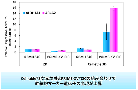
- ※ PRMIE-XV® Cancer Initiating Cells SF Enrichment medium (PRIME-XV® CIC)
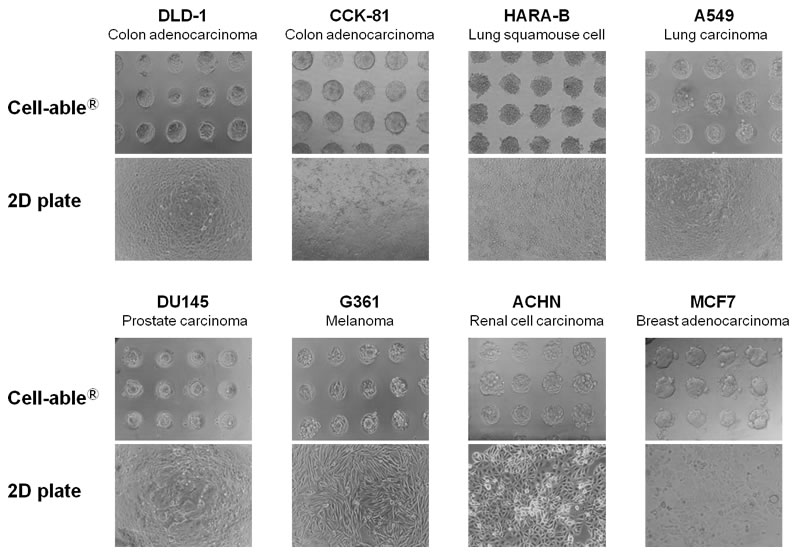
Cell-able® プレートでスフェロイド形成が確認されたがん細胞株は144種類
文献紹介
Cell-able® に関する最近の文献の紹介
【肝細胞培養について(肝毒性、薬物代謝、組織の構造、長期培養、アルブミン産生能)】
- OGIHARA, Takuo, et al.,, Utility of human hepatocyte spheroids for evaluation of hepatotoxicity., Fundamental Toxicological Sciences, 2015, 2. 1: 41-48.
- OHKURA, Takako, et al. Evaluation of human hepatocytes cultured by three-dimensional spheroid systems for drug metabolism. Drug metabolism and pharmacokinetics, 2014, 29. 5: 373-378.
- KUSUMOTO, K., et al. A new high-throughput analysis for drug metabolism profiling using liquid chromatography coupled with tandem mass spectrometry. Drug Res (Stuttg), 2013, 63. 4: 171-176.
- ENOSAWA, Shin, et al. Construction of Artificial Hepatic Lobule-Like Spheroids on a Three-Dimensional Culture Device. Cell Medicine, 2012, 3. 19-23.
- IKEDA, Y., et al. Long-term survival and functional maintenance of hepatocytes by using a microfabricated cell array. Colloids Surf B Biointerfaces, 2012, 97. 97-100.
- MIYAMOTO, Y., et al. Preconditioned cell array optimized for a three-dimensional culture of hepatocytes. Cell Transplant, 2009, 18. 5: 677-681.
- OTSUKA, Hidenori, et al. Two-Dimensional Multiarray Formation of Hepatocyte Spheroids on a Microfabricated PEG-Brush Surface. ChemBioChem, 2004, 5. 850-855.
【肝細胞の分化について】
- WANG, Wenjie, et al. 3D spheroid culture system on micropatterned substrates for improved differentiation efficiency of multipotent mesenchymal stem cells. Biomaterials, 2009, 30. 14: 2705-2715.
【がん研究】
- TAN, Aaron C and KONCZAK, Izabela OS 2-1 Modulatory effects of native Australian fruits polyphenols on carcinogenesis pathways
- WAKAHASHI, Senn, et al. (2013). VAV1 represses E-cadherin expression through the transactivation of Snail and Slug: a potential mechanism for aberrant epithelial to mesenchymal transition in human epithelial ovarian cancer. Translational Research. 162: 181-190. http://dx.doi.org/10.1016/j.trsl.2013.06.005
- MAKINO, Jun, et al. (2015). CRGD-installed polymeric micelles loading platinum anticancer drugs enable cooperative treatment against lymph node metastasis. Journal of Controlled Release. 220: 783-791. http://www.sciencedirect.com/science/article/pii/S0168365915301838
- YOKOTA, Koichi, et al. (2013). Stem-like cell characteristics of cancer spheroids grown in a microfabricated cell array three-dimensional culture system Cell-ableTM Oncology. Cancer research. 73: 3838-3838. http://cancerres.aacrjournals.org/content/73/8_Supplement/3838
- ITAKA, Keiji, et al. (2015). Gene transfection toward spheroid cells on micropatterned culture plates for genetically-modified cell transplantation. JoVE (Journal of Visualized Experiments): e52384-e52384
- PRAVEEN, Kesavan Nair, et al. (2012). Evaluation of Cell-able spheroid culture system for culturing patient derived primary tumor cells. Cancer research. 72: 5270-5270. http://cancerres.aacrjournals.org/content/72/8_Supplement/5270
【その他】
- EDMONDSON, Rasheena, et al. (2014). Three-dimensional cell culture systems and their applications in drug discovery and cell-based biosensors. ASSAY and Drug Development Technologies. 12: 207-218. https://www.ncbi.nlm.nih.gov/pmc/articles/PMC4026212/pdf/adt.2014.573.pdf
- UCHIDA, S., et al. (2014). An injectable spheroid system with genetic modification for cell transplantation therapy. Biomaterials. 35: 2499-2506. http://www.ncbi.nlm.nih.gov/pubmed/24388386 1442.MIYAMOTO, Yoshitaka, et al. (2013). Observation of Positively Charged Magnetic Nanoparticles Inside HepG2 Spheroids Using Electron Microscopy. Cell Medicine. 5: 89-96. https://www.ncbi.nlm.nih.gov/pmc/articles/PMC4733861/pdf/CM-5-89.pdf
- FURUHATA, Yuichi, et al. (2016). Small spheroids of adipose‐derived stem cells with time‐dependent enhancement of IL‐8 and VEGF‐A secretion. Genes to Cells. 21: 1380-1386. http://onlinelibrary.wiley.com/doi/10.1111/gtc.12448/abstract
- MIYAMOTO, Y, et al. (2012). Polysaccharide functionalized magnetic nanoparticles for cell labeling and tracking: A new three-dimensional cell-array system for toxicity testing. Nanomaterials for Biomedicine, ACS Publications: 191-208
- UCHIDA, Satoshi, et al. (2016). Treatment of spinal cord injury by an advanced cell transplantation technology using brain-derived neurotrophic factor-transfected mesenchymal stem cell spheroids. Biomaterials. 109: 1-11. http://www.sciencedirect.com/science/article/pii/S0142961216304859
- UCHIDA, S., et al. An injectable spheroid system with genetic modification for cell transplantation therapy. Biomaterials, 2014, 35. 8: 2499-2506.
心毒性評価に関する学会発表
- 第43回日本毒性学会学術年会 (2016)
ヒトiPS心筋を用いたマルチスフェロイドCaイメージングによる化合物催不整脈性HTS評価システム
長倉廷1, 松原孝宜2, 澤田光平1
1) エーザイ株式会社筑波研究所グローバルCV 評価部, 2) 横河電機株式会社ライフサイエンスセンター
- 第43回日本毒性学会学術年会 (2016)
ヒトiPS由来心筋細胞を用いたマルチスフェロイドイメージング解析による抗がん剤の心毒性評価に関する検討
Evaluation study of cardiotoxicity of anti-cancer drugs using multi-spheroid imaging analysis of human induced pluripotent stem cell-derived cardiomyocyte
近藤卓也,一ツ町裕子,一ツ町知明,蟹江尚平,平野隆之,森田文雄,箱井加津雄,大鵬薬品工業株式会社
Takuya KONDO, Hiroko HITOTSUMACHI, Tomoaki HITOTSUMACHI, Shohei KANIE, Takayuki HIRANO, Fumio MORITA, Kazuo HAKOI, Taiho Pharmaceutical Co., Ltd.
- 15th World Preclinical Congress 2016
Multi-Spheroid Imaging Analysis of Human iPS Cell-Derived Cardiomyocyte as a Assessment Model for the Acute Effects of Cardiotoxicity of Anti-Cancer Drugs
Takuya, Kondo, Taiho Pharmaceutical Co., Ltd.
- ㈶バイオインダストリー協会"未来へのバイオ技術" 勉強会 (2016)
「ハイコンテントアナリシス(HCA)技術の進化」iCell心筋細胞を用いたマルチスフェロイドイメージング解析による新たな心毒性評価法の開発」
長倉廷(エーザイ株式会社 バイオファーマシューティカル・アセスメント機能ユニットグローバルCV評価部)
- 第6回学術年会日本安全性薬理研究会 (2015)
Multi-spheroid imaging analysis of human induced pluripotent stem cell-derived cardiomyocyte by Cellvoyager CV7000 system for the assessment of in vitro caridiotoxicity
TadashiNAGAKURA1), Takayoshi MATSUBARA2), Ko ZUSHIDA3) and Kohei SAWADA1)
1) Global CV Assessment Unit, Tsukuba Research Laboratory, Eisai Co., Ltd.,2) Engineering Team, Life Science Business HQ, Yokogawa Electric corporation,3) Product Development & Operations, iPS Portal, Inc.
- 第42回日本毒性学会学術年会 (2015)
ヒトiPS由来心筋細胞を用いたマルチスフェロイドイメージング解析の in vitro心毒性評価系としての最適化検討
長倉廷1, 松原孝宜2, 圖子田康3, 澤田光平1
1) エーザイ株式会社筑波研究所グローバルCV 評価部, 2) 横河電機株式会社ライフサイエンスセンター営業部営業技術課, 3) 株式会社iPS ポータルプロダクト開発事業部
- Cellular Dynamics International, iForum 2015
A Novel Method for the Assessment of Cardiotoxicity by Multi-spheroid Imaging Analysis of Human Induced Pluripotent Stem Cell-derived Cardiomyocytes
Kohei Sawada, PhD, Eisai Co., Ltd.
関連製品
トピックス すべてを見る
- 2025/01/28 製品情報 Cell-able®製品販売終了のお知らせ
- 2025/01/27 メルマガ S-BIO Insight『毛包誘導形成にPrimeSurfaceを活用!最新研究の成果をご報告』
- 2025/01/20 製品情報 新製品「EZGlyco® mAb-N kit with APTS」発売のお知らせ
- 2025/01/20 製品情報 ステムフル®の新規アプリケーションデータ「細胞回収用低吸着遠沈管ステムフル®のF-hiSIEC™(ヒトiPS細胞由来腸管上皮細胞)回収における比較試験」を追加しました
- 2024/12/25 メルマガ S-BIO Insight『ヒト母乳の糖鎖分析による新たな発見』を配信しました
- 2024/12/13 製品情報 PrimeSurafce®の新規アプリケーションデータ「PrimeSurface®プレート96Vを使用した階層スフェロイド型BBBモデル」を追加しました


